Contact us
Send Us Your Details
Please fill out the form below and a member of our team will be in contact with you.


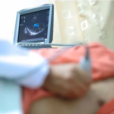
Echocardiography, a non-invasive imaging technique, has revolutionized the field of cardiology by providing detailed insights into the structure and function of the heart. In this comprehensive exploration, we delve into the fundamentals of echocardiography, its various types, applications, and its invaluable role in cardiovascular diagnosis and management.
Echocardiography, often called an “echo,” employs sound waves to create detailed images of the heart’s chambers, valves, and surrounding structures. It is a pivotal diagnostic tool that allows healthcare providers to assess cardiac function, identify abnormalities, and guide treatment decisions.
Types of Echocardiography: Tailoring Imaging to Clinical Needs
Echocardiography is generally a painless and non-invasive procedure. During a standard TTE, the patient lies on their left side, and a small amount of gel is applied to the chest. The transducer is then moved across the chest to capture images, and the entire procedure typically takes about 30-60 minutes. TEE involves a specialized probe, and while it may be slightly more uncomfortable, it is usually well-tolerated.
Interpreting echocardiographic images requires specialized skills and expertise. Cardiologists analyze the size and thickness of heart chambers, the movement of heart valves, the efficiency of blood ejection, and other subtle details. Echocardiography allows for identifying conditions at their early stages, enabling timely intervention and management.
Echocardiography is not confined to adults but is crucial in pediatrics. Pediatric cardiologists use echocardiography to assess congenital heart defects, evaluate cardiac function in children with various diseases, and monitor the effectiveness of treatments. The non-invasive nature of echocardiography is particularly advantageous in the delicate realm of pediatric care.
Challenges and Considerations: Navigating Diagnostic Limitations
While echocardiography is a powerful diagnostic tool, it does have limitations. Obesity, chronic lung disease, and certain chest deformities can affect image quality. Additionally, echocardiography may not provide detailed views of specific structures, necessitating complementary imaging modalities when required.
Echocardiography stands as a cornerstone in cardiovascular diagnostics, offering a window into the intricate workings of the heart. Its versatility, non-invasive nature, and ability to provide real-time insights make it an invaluable tool for healthcare providers. From detecting structural anomalies to guiding intricate cardiac procedures, echocardiography empowers cardiovascular care, providing more accurate diagnoses and tailored treatment strategies. As technology advances, the future promises even more refined and detailed cardiac imaging, further elevating the standard of cardiovascular medicine.


Disclaimer:
The information on this website is provided for educational and information purposes only and is not medical advice. Always consult with a licensed medical provider and follow their recommendations regardless of what you read on this website. If you think you are having a medical emergency, dial 911 or go to the nearest emergency room. Links to other third-party websites are provided for your convenience only. If you decide to access any of the third-party websites, you do so entirely at your own risk and subject to the terms of use for those websites. Neither The Cardiac and Vascular Interventional Group, nor any contributor to this website, makes any representation, express or implied, regarding the information provided on this website or any information you may access on a third-party website using a link. Use of this website does not establish a doctor-patient relationship. If you would like to request an appointment with a health care provider, please call our office at (469) 437-3560.